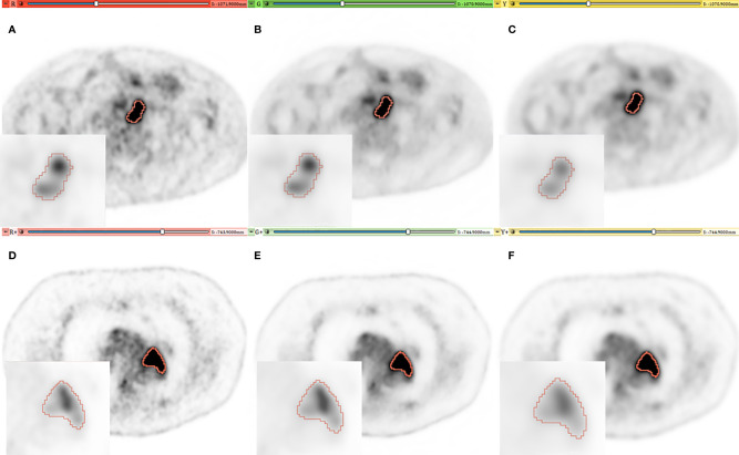
Figure 1.
Representative PET imaging of two lesions in different patients with a SUV windowing of (0–5). (A, D) [red], (B, E) [green], (C, F) [yellow] for original, AI and EARL images respectively. A Zoom is added on each image with SUV windowing between (0–25) and (0–30) for the first and second patient.

