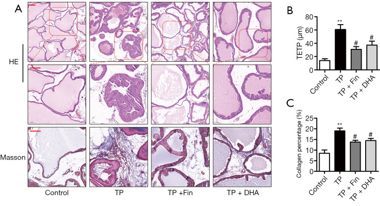
Figure 2.
The effects of dihydroartemisinin on the histological changes and collagen deposition in rats. (A) H&E staining of prostate tissues (top panel: 100× magnification, bar =50 µm; second panel: 200 × magnification, bar =50 µm). Masson’s trichrome staining of prostate tissues (third panel: 200× magnification, bar =50 µm). (B) The thickness of epithelium tissue from prostate (TETP) (n=4; **P<0.01 versus Control group; #P<0.05 versus TP group). (C) Quantification of Masson’s trichrome staining. The percentage area of collagen fiber was quantified from three random fields of view (200× magnification) from each tissue slices (n=4; **P<0.01 versus Control group; #P<0.05 versus TP group). H&E, hematoxylin and eosin; DHA, dihydroartemisinin; TP, testosterone propionate.

