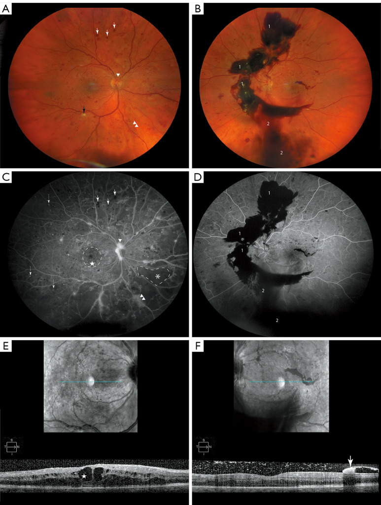
Figure 1.
Features of DR. (A) Color fundus photo of the right eye of a patient with PDR, highlight pathologic changes which include ischemic damage to nerve fibers known as “cotton wool” spot (black arrow), dot-blot hemorrhages (white arrows), NVD (arrowhead) and NVE (double arrowhead). (B) The left eye of the same patient with PDR denotes advanced disease progression, highlighted are pathologic features including pre-retinal/sub-hyaloid bleeding [1] and VH [2]. (C,D) Corresponding FFA images. Pathological features shown here include MAs (dashed arrow) with corresponding features previously mentioned (A,B) and respectively denoted. Petaloid pattern of CME is encircled with dashed line. Area of capillary dropout is marked with asterix (*) and demarcated with dashed line. (E) OCT image of right eye shows cystoid macular edema CME that is more prominent in central macula (star). (F) OCT image of left eye. Hyper-reflective lesion (white arrow) representing pre-retinal/sub-hyoid hemorrhage. DR, diabetic retinopathy; PDR, proliferative diabetic retinopathy; NVD, neovascularization of the disc; NVE, neovascularization elsewhere; FFA, fundus fluorescein angiogram; MA, microaneurysm; VH, vitreous hemorrhage; CME, cystoid macular edema; OCT, optical coherence tomography.

