
Figure 1. ACM patients show high plasma oxLDL, cardiac lipid peroxidation, and oxLDL receptor levels.
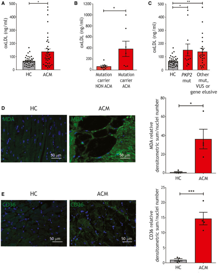
- oxLDL plasma concentration in ACM patients and HC (n = 36; Mann–Whitney test).
- oxLDL plasma concentration in mutated ACM (n = 7) and non‐ACM relatives, carriers of the same causative mutation (n = 9; Mann–Whitney test).
- oxLDL plasma concentration in ACM patients carriers of a PKP2 mutation and ACM patients carriers of other desmosomal or non desmosomal mutations, or gene elusive (n = 10 vs. n = 26; one‐way ANOVA).
- Representative images of MDA immunostaining (green) on ACM and HC ventricular tissue sections and relative quantification (n = 4 biological replicates; two‐tailed Student’s t‐test). Nuclei are counterstained with Hoechst33342 (blue).
- Representative images of CD36 immunostaining (green) on ACM and HC ventricular tissue sections and relative quantification (n = 4 biological replicates; two‐tailed Student’s t‐test). Nuclei are counterstained with Hoechst33342 (blue).
Data information: mean ± SEM. *P < 0.05; **P < 0.01; and ***P < 0.001.
Source data are available online for this figure.