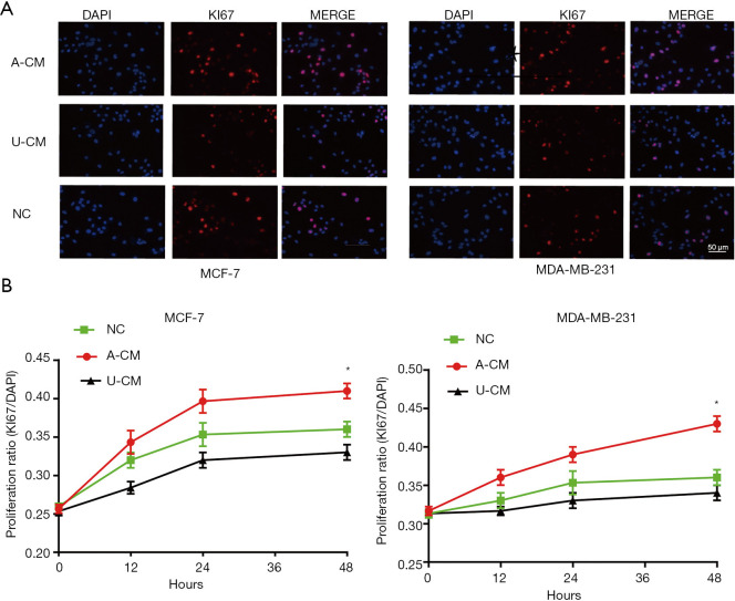
Figure 2.
Immunofluorescence assay shows the proportion of Ki67-positive cells were significantly higher in the A-CM group. (A) Representative fluorescent picture of MCF-7 cells and MDA-MB-231 cells at 48 h of incubation with A-CM or U-CM (upper; magnification, 400×). (B) Corresponding quantification using the blue mean fluorescence intensity of DAPI at 12, 24, and 48 h of incubation with A-CM or U-CM (lower). (* indicates P<0.05; NC: means natural control group, which were treated with free DMEM/F12). ADSCs, adipose-derived stem cells; UMSCs, umbilical mesenchymal stem cells; A-CM, ADSCs-related medium; U-CM, UMSCs-related medium; DAPI, 4',6'-diamidino-2-phenylindole hydrochloride; DMEM, Dulbecco’s modified Eagle’s medium.

