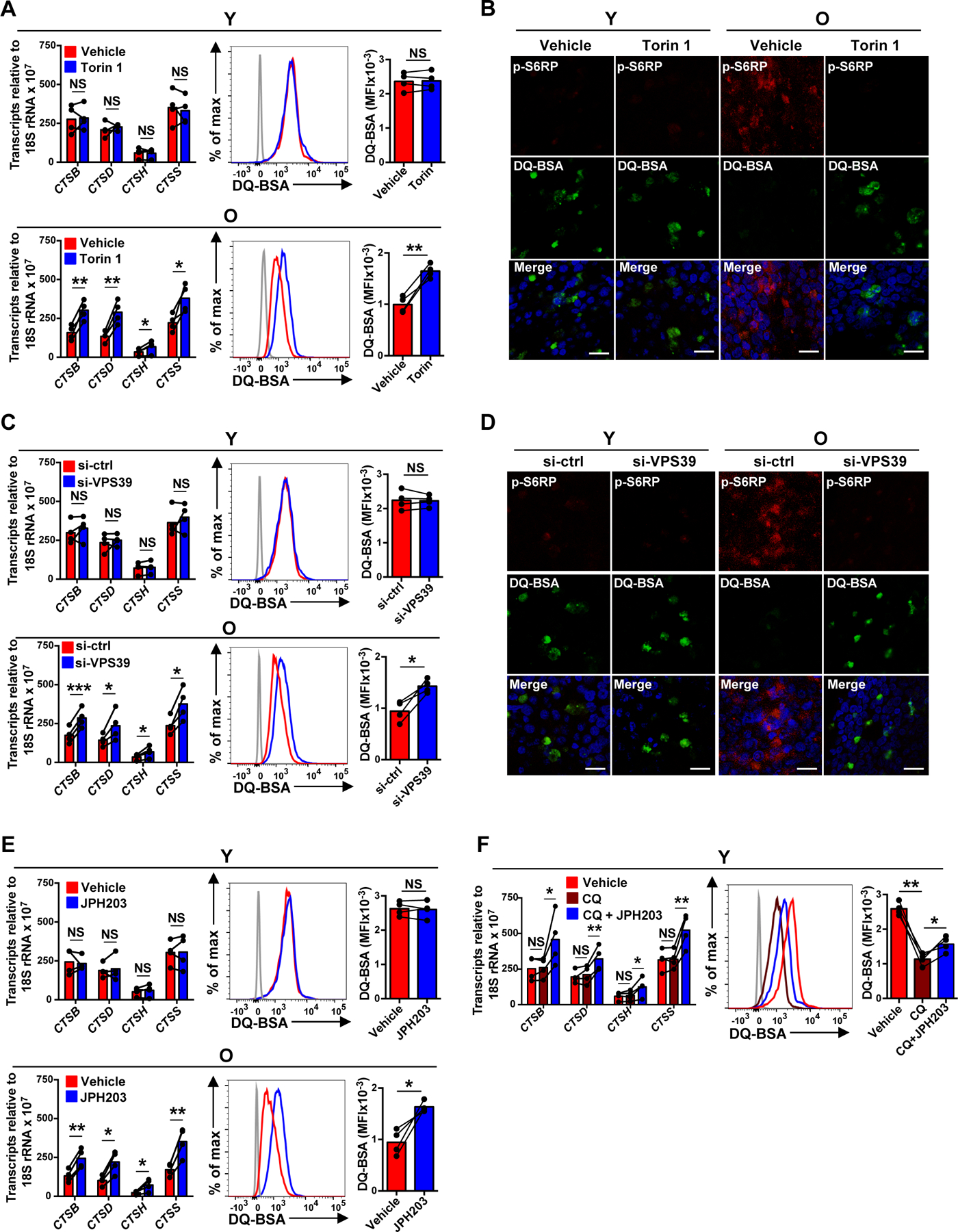
Fig. 3. Sustained activation of late endosomal mTORC1 suppresses lysosomal activities in naïve CD4+ T cell responses.

(A–F) Naïve CD4+ T cells from young (Y) and older (O) individuals were activated for 5 days. Indicated inhibitor or vehicle control was added for the last 2 days of culture (A–B and E–F). Alternatively, cells were transfected with control or VPS39 siRNA and then activated with anti-CD3/anti-CD28 beads for 5 days (C–D). (A, C, E and F) Lysosomal cathepsins expressions were determined by qRT-PCR (left); results are normalized to control samples using 18S rRNA as internal control; mean ± SD of four experiments. Lysosomal activities were determined by flow cytometry-based analysis of cells treated with 5 μg/mL of DQ-BSA for 6 hours. Results are shown as representative histograms (middle) and cumulative data from four experiments (right). The gray histogram represents DQ-BSA free samples. (B and D) Cells were treated with DQ-BSA (green) and stained with anti-pS6RP (red). Confocal images representative of two independent experiments show an inverse relationship between mTORC1 and lysosomal activity. Scale bar, 20 μm. Comparison by two-tailed paired t test (A, C, E and F). *p < 0.05, **p < 0.01; NS, not significant.
