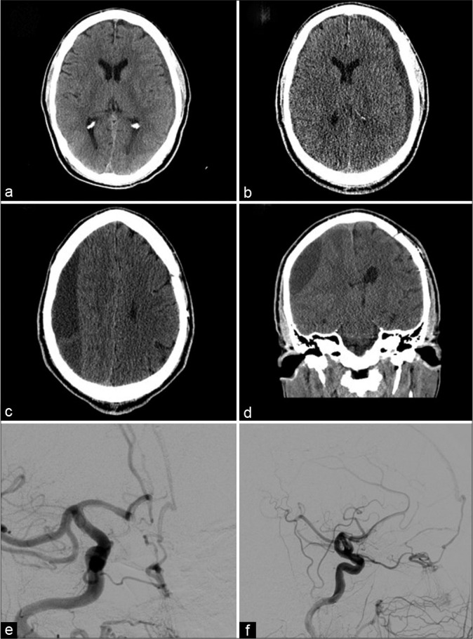
Figure 3:

(a) Axial noncontrast CT demonstrates 5 mm left convexity chronic subdural hematoma corresponding to the patient’s initial presentation. (b) Axial noncontrast CT demonstrates interval increase in size of the left convexity subdural hematoma. (c and d) Axial and coronal noncontrast CT performed 1 month later demonstrates a new large right subacute on chronic subdural hematoma. (e and f) Anteroposterior and lateral views, selective catheter angiogram, and right common carotid artery injection demonstrating a cribriform plate dural arteriovenous fistula with ophthalmic artery feeders draining into a right frontal cortical vein.
