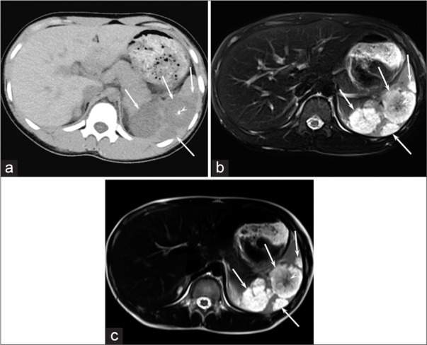
Figure 1:

A 15-year-old girl with GLA (Patient 3). (a) Unenhanced CT image shows hypodense focal splenic lesions larger than 10 mm (arrows) with central calcification (arrowhead). (b and c) Respiratory-triggered fat-suppressed T2-weighted fast spin-echo (TR/TE, 1,600/80 ms) (b) and T2-weighted single-shot fast spin-echo (TR/TE, 12,146/150 ms) (c) images show hyperintense focal splenic lesions larger than 10 mm (arrows) with central hypointense areas (arrow heads).
