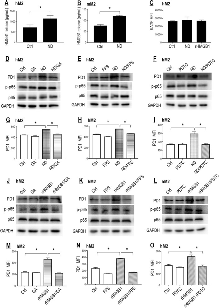
Fig. 5.
Nano-DOX induced PD-1 in TAMs through activation of the HMGB1/ RAGE/NF-κB pathway. A, B Nano-DOX stimulated HNGB1 secretion from the hM2 and mM2. C Neither Nano-DOX nor HMGB1 induced RAGE in the hM2. D–I Pharmacological blocking of the HMGB1/RAGE/NF-κB pathway suppressed PD-1 induction by Nano-DOX in the hM2. J–O HMGB1 induced PD-1 in the hM2, which was repressed by blocking of the HMGB1/RAGE/NF-κB pathway. Cell surface PD-1 and RAGE were assayed by FACS analysis of immunofluorescent staining and protein levels thereof were assayed by western blotting. FACS histogram geometric means were used to quantify mean fluorescence intensity (MFI). Values were means ± SD (n = 3, *p < 0.05). Glycyrrhizic acid (GA) both neutralizes HMGB1’s cytokine activity and suppresses its secretion. FPS-ZM1 is a high-affinity inhibitor of RAGE. Pyrrolidine dithiocarbamate (PDTC) is a selective inhibitor of NF-κB. Drug concentration was 2 μg/mL for DOX and Nano-DOX and HMGB1 (0.5 μg/mL for hM2 and 2 μg/mL for the A549 cells) in the in vitro experiments and treatment duration was 24 h. Representative FACS dot plots for C, G–I and M–O were provided in Additional file 1: Figure S5

