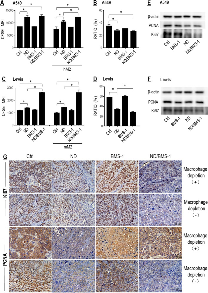
Fig. 8.
BMS-1 and Nano-DOX synergistically inhibited proliferation of lung cancer cells. A BMS-1 enhanced Nano-DOX’s action to inhibit A549 cell growth only in mixed culture with the hM2. B Proportion of A549 cells in the mixed culture. C BMS-1 enhanced Nano-DOX’s action to inhibit Lewis cell growth both in single culture and in co-culture with the mM2. D Proportion of Lewis cells in the mixed culture. E, F Effects of Nano-DOX, BMS-1 and the combination thereof on the protein levels Ki67 and PCNA in the in vitro A549 and Lewis cells. G Nano-DOX treatment led to decreased immunohistological staining of Ki67 and PCNA in subcutaneous xenografts of Lewis cells in mice. Cell growth was assayed by FACS analysis of decay of CFSE staining. Ki67 and PCNA protein was assayed by western blotting. FACS histogram geometric means were used to quantify mean fluorescence intensity (MFI). Values were means ± SD (n = 3, *p < 0.05). Drug concentration was 2 μg/mL for DOX and Nano-DOX and 1 μM for BMS-1 in the in vitro experiments and treatment duration was 24 h. Representative FACS zebra plots for A–D were provided in Additional file 1: Figure S8

