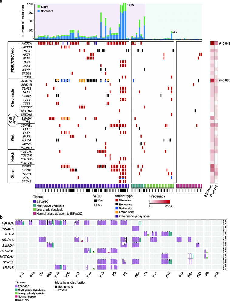
Fig. 2.
Mutational landscape of EBVaGCs, dysplasia samples, and normal tissues. a Top: the number of silent and non-silent mutations of each sample. Middle: somatic mutations (SNVs and INDELs) of EBVaGC-associated genes in different pathways. Genes are ranked by their mutated frequencies. Bottom: histological types and whole-genome doubling (WGD) status of each sample are indicated in different colors. Right: heatmap comparing mutated frequencies of each gene (row) in EBVaGCs and their precursor lesions over patients. The statistical significance is shown (Fisher’s exact test). b Bar plots showing the cancer cell fraction (CCF) of each mutation in recurrently mutated genes. Solid bars denote the mutations that are shared by multiple samples from the same patient. Hollow bars denote the mutations that are private in single samples. It should be noted that there existed multiple mutations in a specific gene within one sample, and these mutations are vertically stacked with an adjusted scale of CCF values. Different colors indicate different histological types. The standard deviation is indicated

