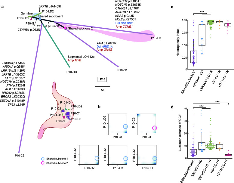
Fig. 4.
Phylogenetic relationships of multiples samples of representative patients. a Top: phylogenetic tree for patient P10. The length of each line is proportional to the number of mutations and copy number alterations (CNAs). Gray lines represent the clonal mutations shared by multiple samples. A subclone in one LD sample of P10 (P10-LD2) presenting as the common ancestor of all other histological advanced samples of P10, and another subclone shared by two EBVaGCs (P10-C1 and P10-C3) are indicated. The shaded area contains mutations present in the shared subclone on the corresponding branches. Bottom: geographical locations of all samples in patient P10. Histological types of all samples are indicated in different colors. b Two-dimensional density plots showing the CCF distribution of pairwise samples in P10. c Box plots depicting the heterogeneity index (HI) of pairwise samples in each patient. All pairwise samples are divided into 5 groups. Wilcoxon rank sum test, ***P < 0.001. d Box plots depicting the Euclidean distance of CCF of pairwise samples in each patient. Wilcoxon rank sum test, ***P < 0.001

