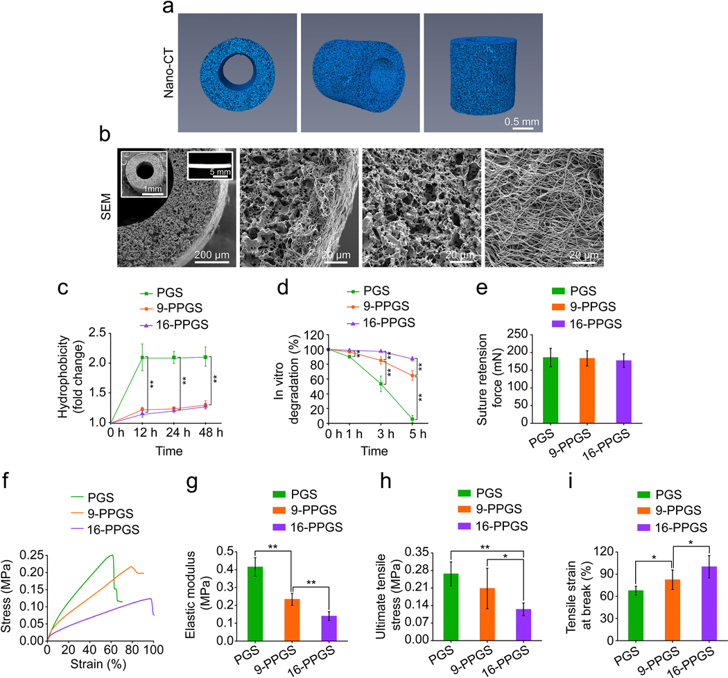
Fig. 1.
(a) Reconstructed 3D images of nano-CT scanning of grafts. (b) SEM images of grafts. Insets: low magnification SEM images of the graft (left) and macroscopic view of the graft (right). (c) Hydrophobicity of three different types of grafts. (d) In vitro degradation of three different types of grafts. (e) Suture retention forces of three different types of grafts. (f) Representative stress-strain curves of three different types of grafts. (g) Elastic modulus of three different types of grafts. (h) Ultimate tensile stress of three different types of grafts. (i) Tensile strain at break of three different types of grafts. * indicates p < 0.05, compared between two groups; ** indicates p < 0.01, compared between two groups.

