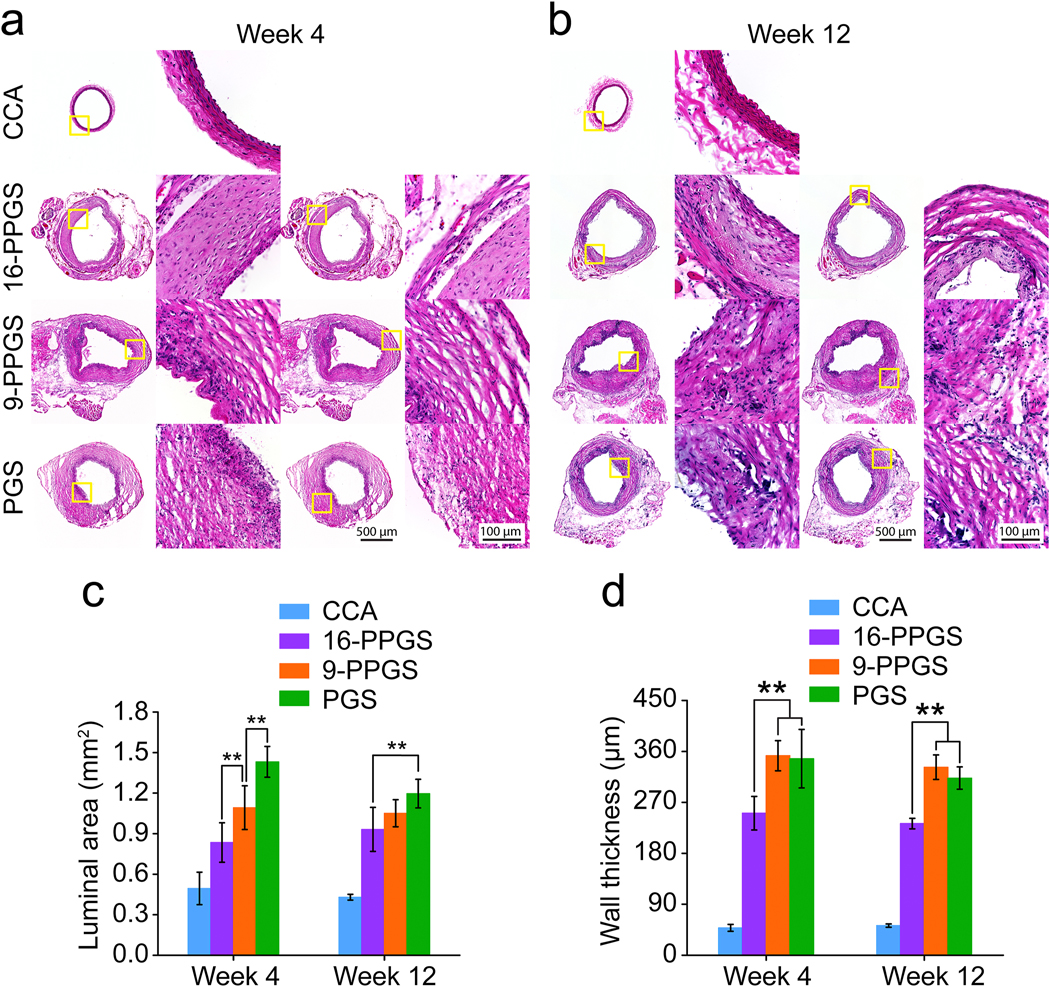
Fig. 4.

(a) H&E staining of three different types of grafts explanted in week 4. (b) H&E staining of three different types of grafts explanted in week 12. (c) Quantification of luminal areas of three different types of grafts explanted in week 4 and 12. (d) Quantification of graft wall thickness of three different types of grafts explanted in week 4 and 12. ** indicates p < 0.01, compared between two groups.
