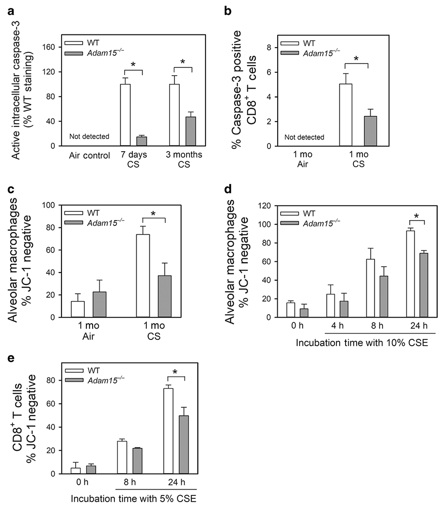
Figure 7: Deficiency of Adam15 in CD8+ T cells and macrophages protects the cells from CS-induced activation of the mitochondrial apoptosis pathway in vivo and in vitro.

A: Alveolar macrophages (AMs) were isolated from the lungs of WT and Adam15−/− mice that had been exposed to air or CS for 1 week or 3 months (Mo) using BAL. B: CD8+ T cells were isolated from the lungs of WT and Adam15−/− mice exposed to air or CS for 1 month as described in Methods. In A and B, immediately after the AMs and CD8+ T cells were isolated they were fixed and then immunostained for intracellular active caspase-3. Staining was quantified, as described in the Methods. Active caspase-3 was not detected in macrophages or CD8+ T cells isolated from air-exposed WT or Adam15−/− mice. Data are mean ± SEM; n = 5 mice/group in A, and n = 4-5 mice/group in B. Data were analyzed using a One-Way ANOVA followed by pair-wise testing with two-tailed Student’s t-tests. *, P < 0.05 versus the group indicated. C: AMs were isolated from the lungs of WT and Adam15−/− mice exposed to air or CS for 1 month using BAL. Activation of the mitochondrial apoptosis pathway was measured as loss of mitochondrial membrane potential (lack of staining of the cells with the mitochondrial dye, JC-1 which stains only viable mitochondria). Data are mean ± SEM; n = 5 mice/group. Data were analyzed using a One-Way ANOVA followed by pair-wise testing with two-tailed Student’s t-tests. *, P < 0.05 versus the group indicated. D: AMs isolated from naïve WT and Adam15−/− mice were incubated at 37°C for up to 24 h with or without 10% CS extract (CSE). E: CD8+ T cells isolated from spleen of naïve WT Adam15−/− mice were incubated at 37°C for up to 24 h with or without 5% CSE. In D and E, activation of the mitochondrial apoptosis pathway was measured as loss of mitochondrial membrane potential (lack of staining of the cells with JC-1). Data are mean ± SEM; n = 3 separate experiments in D and E. Data were analyzed using a One-Way ANOVA followed by pair-wise testing with two-tailed Student’s t-tests. *, P < 0.05 versus the group indicated.
