Figure 2.
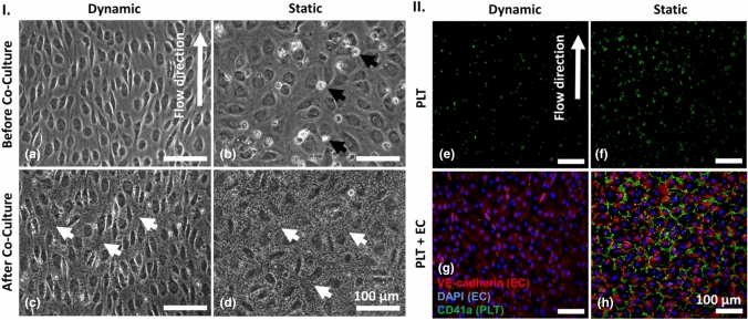
Endothelial cells (EC), platelets (PLT) and co-cultures of EC and PLT under dynamic and quasi-static conditions. (I) Phase contrast images: EC after six days of cultivation under laminar flow (10 dyn cm−2, a) or quasi-static conditions (0.01 dyn cm−2, b) prior to co-culture (black arrows indicate dead EC); EC and PLT (visible here as small, granular speckles indicated by white arrows) after 1 h of co-culture under laminar flow (c) or quasi-static conditions (d). (II) Immunostaining: PLT (CD41a-FITC-labeled, green) cultivated in the absence of EC under laminar flow (e) or quasi-static conditions (f); Co-cultures of EC (VE-cadherin-Alexa Fluor 555- and DAPI-labeled, red with blue nuclei) and PLT after 1 h under laminar flow (g) or quasi-static conditions (h).

