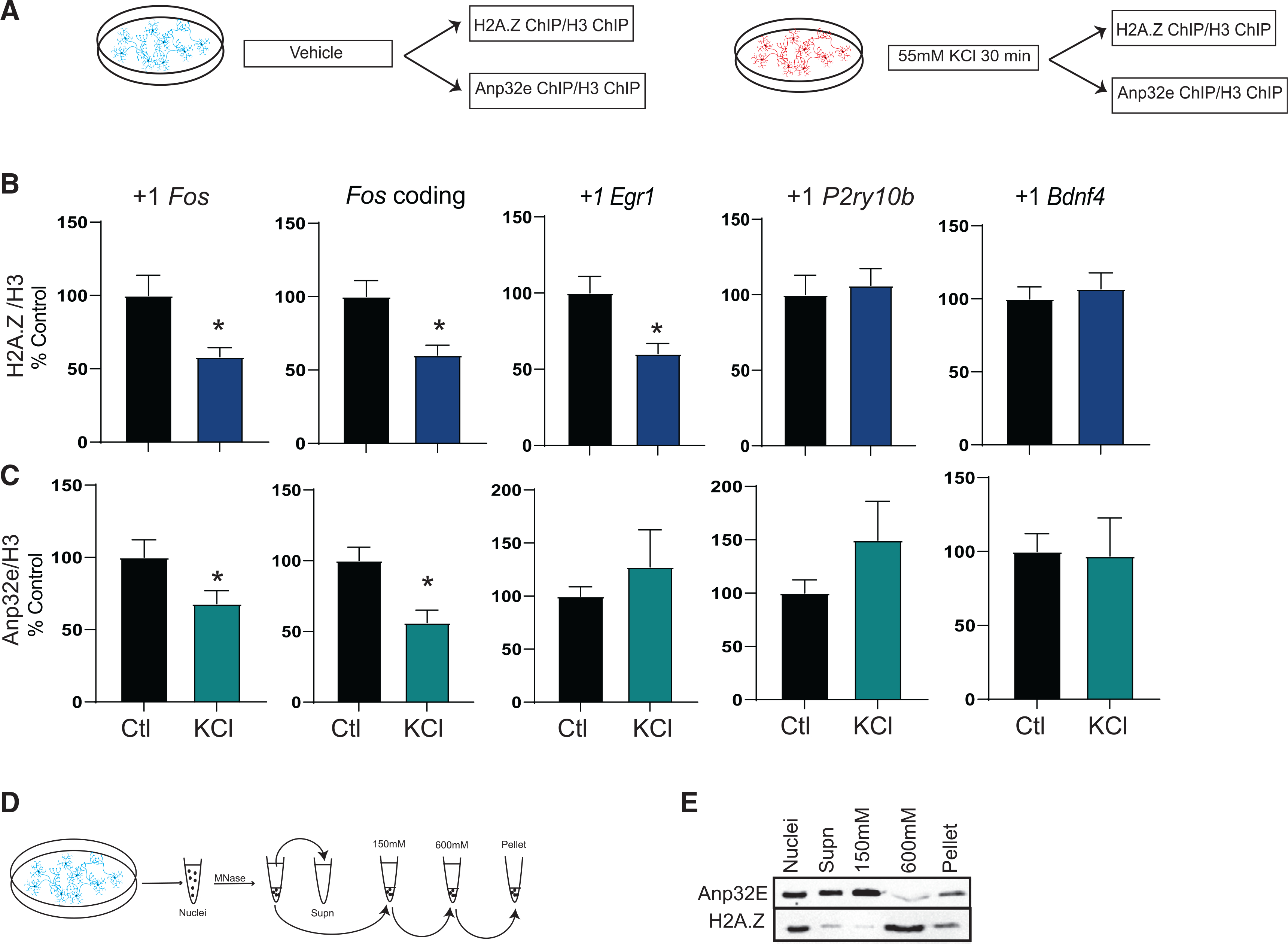
Figure 1. H2A.Z and Anp32e binding is reduced with neural depolarization.

(A) Schematic representation of the experimental workflow in cultured hippocampal neurons.
(B and C) Chromatin immunoprecipitation (ChIP) was used to assess binding of (B) H2A.Z (n = 13–17/group) or (C) Anp32e (N = 8–10/group) in the first nucleosome downstream (+1 nucleosome) of the transcription start site (TSS) and in the coding region 30 min after KCl depolarization. H2A.Z binding is normalized to H3 and data are expressed as mean ± SEM. *p ≤ 0.05.
(D) Schematic representation of nucleosome salt fractionation.
(E) MNase fractionation and salt extraction of primary hippocampal neuronal nuclei were used to assess Anp32e binding to H2A.Z in distinct chromatin fractions.
