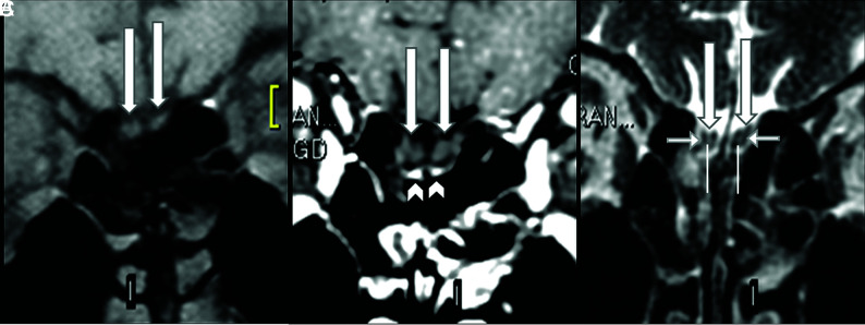
FIG 1.
Normal olfactory bulbs and susceptibility artifacts seen on 1.5T MR imaging before the COVID-19 pandemic. The coronal precontrast fat-suppressed T1WI (A) and the postcontrast (B) and coronal FSE T2WI (C) demonstrate normal olfactory bulbs (long arrows). The olfactory bulbs are isointense to the cerebral cortex and normally hypointense on pre- (A) and postgadolinium sequences (B) and do not enhance. Susceptibility artifacts on the cribriform plate (B, arrowheads) are bilateral and symmetric below the olfactory bulbs and do not hinder the analysis. On thin-sliced coronal FSE T2WI, the normal olfactory bulbs show a “sandwich-like pattern,” which consists of a hyperintense central area (C, superior extremity of the vertical lines), similar to the cortical gray matter, and a hypointense periphery (C, short horizontal arrows), similar to the white matter that looks like the laminar layers of olfactory bulbs on histology.

