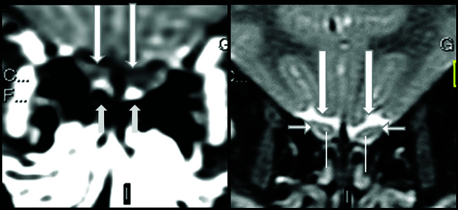
FIG 2.
Normal olfactory bulbs and susceptibility artifacts seen on 1.5T MR imaging before the COVID-19 pandemic. The olfactory bulbus are normal, being hypointense, and do not enhance on thin-sliced coronal fat-suppressed postcontrast T1WI (A, long arrows). Susceptibility artifacts are bilateral and symmetric and can be recognized as hyperintensity located outside and adjacent to the inferior periphery of the normal olfactory bulbs, mainly at cribriform plate (A, short arrows). On coronal T2WI, the normal sandwich-like pattern is observed as the central portion of olfactory bulbs showing hyperintensity (B, superior extremity of the line), similar to that of gray matter, and the peripheral portion showing hypointensity, similar to that of white matter (B, short horizontal arrows).

