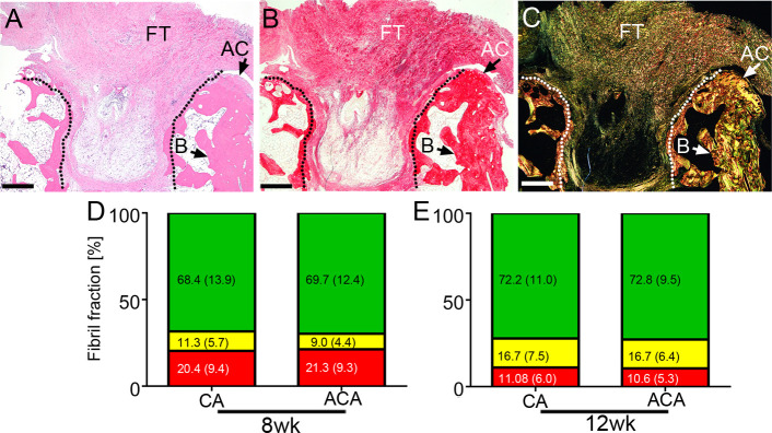
Fig 5. A histological assay of the pannus-like scar tissue formed in an osteochondral defect (delineated).
The site of injury was stained with H&E (A) and with collagen-specific picrosirius red (B&C). The picrosirius red-stained samples were observed in normal light (B) and polarized light (C). Fibrotic tissue (FT), bone (B), and fragments of the articular cartilage (AC) are indicated. D&E, A summary of measurement of the percentages of the green birefringence (GB) fibrils, yellow birefringence (YB) fibrils, and red birefringence (RB) fibrils formed in the CA-treated and the ACA-treated rabbits. Corresponding segments of the bars include the means and standard deviations (in parentheses). Data for the 8wk and the 12wk groups are presented. Bars = 1 mm.

