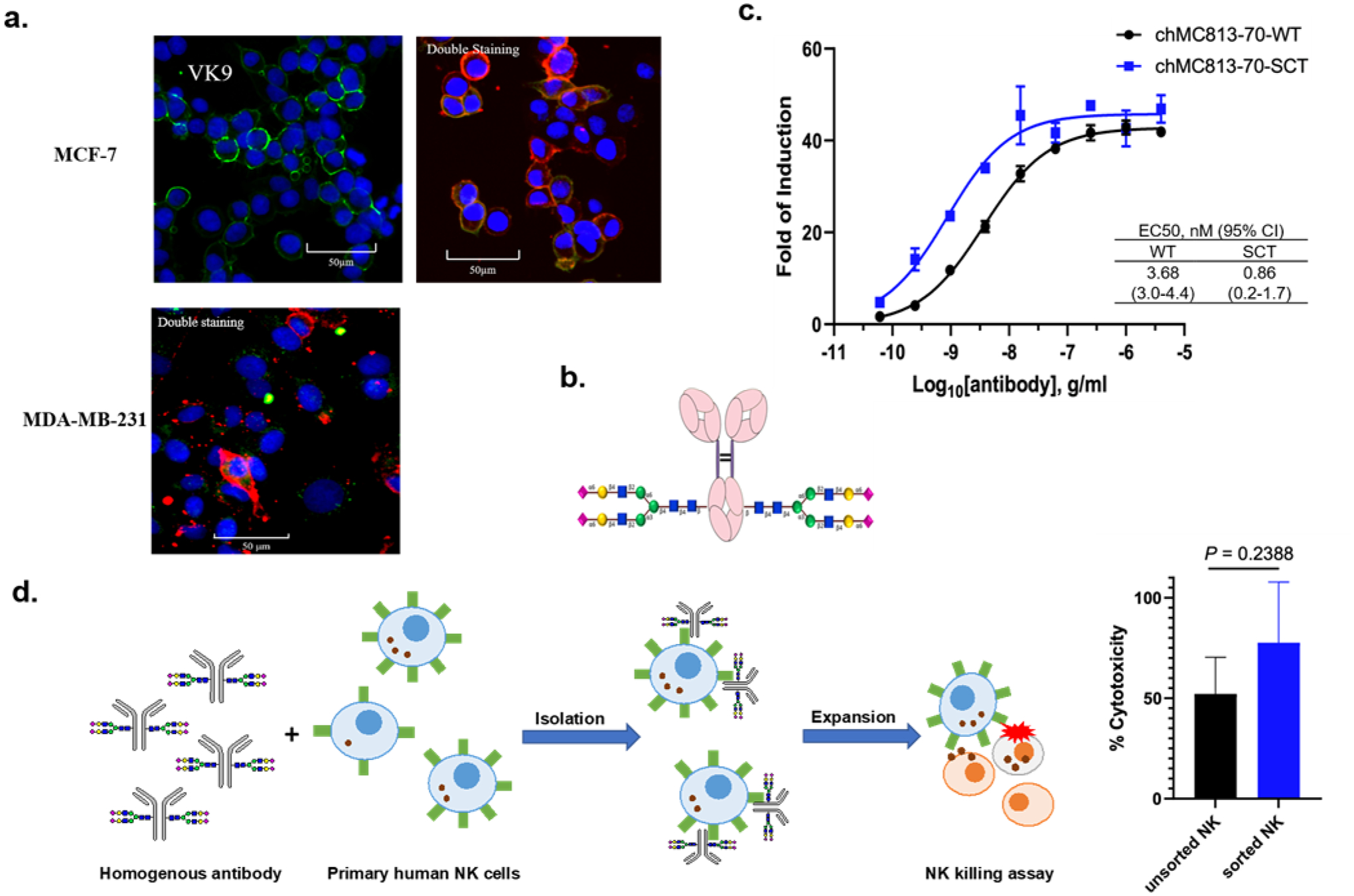
Figure 7.

a. Opera Phenix™ Image of MCF-7 and MDA-MB-231 breast cancer cells stained with FICT-conjugated VK9 (green, targeting Globo-H) only, or with the mixture of APC-conjugated MC813–70 (red, targeting SSEA4) and FICT-conjugated VK9 in a 1:50 ratio (0.5 μg/ml : 25 μg/ml). The results showed that cells are still predominantly interacting with MC813–70. At 1:1 ratio, the cancer cell is completely occupied by MC813–70. Nucleus is in blue (Hoechst 33342 (x 1/500). b. Homogeneous antibody MC813–70-SCT with maximized ADCC activity for the isolation of NK cells enriched with FcγIIIA receptor responsible for the ADCC activity. c. Comparison of heterogeneous and homogeneous chMC813–70 in ADCC assay. The half-maximal effective concentrations (EC50) for chMC813–70-WT were 3.68 ng/mL against MDA-MB-231, and chMC813–70-SCT were 0.86 ng/mL against MDA-MB-231. d. Use of homogeneous antibody chMC813–70-SCT for isolation and expansion of a subpopulation of NK cells enriched with FcγIIA receptor which exhibited around 23% increase in killing the target cells (breast cancer cell line MDA-MB-231 with high expression of SSEA4).
