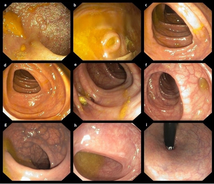
Figure 5. Lower gastrointestinal colonoscopy.
A lower gastrointestinal colonoscopy shows the ileum (a), cecum (b), and ascending and transverse colon (c, d) with no obvious pathology. There is diverticulosis of the descending and sigmoid colon (e, f), rectum with phlebectasia (g, h), and the anal canal (i) with no pathology.

