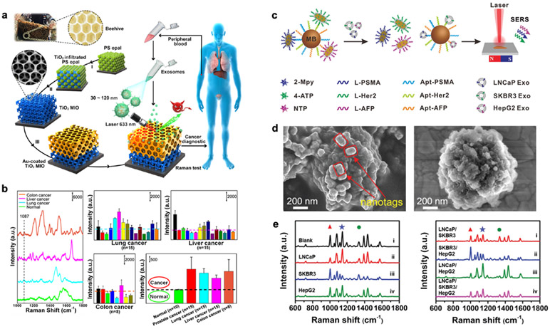
Figure 7. SERS platform.
a Detection process and design inspiration of the Au-coated TiO2 MIO SERS substrate. b SERS spectra of exosomes separated from plasma of different cancer patients and normal individuals. SERS intensity at 1,087 cm−1 is used for analysis. Reprinted with permission from ref. 47. Copyright 2020 American Chemical Society. c Multiplex exosome detection using SERS nanoprobes. Multiplexed capture probes were constructed by the co-modification of three types of aptamer DNAs. Followed by the recognition between aptamers and target exosomes, the SERS probes are released, and SERS signals are attenuated. d SEM images of the hybridized complexes of SERS probes–magnetic bead, and exosome-magnetic bead. e SERS spectra of the complexes obtained in the presence of single (left) and multiple (right) exosomes. Reprinted with permission from ref. 51. Copyright 2020 Royal Society of Chemistry.

