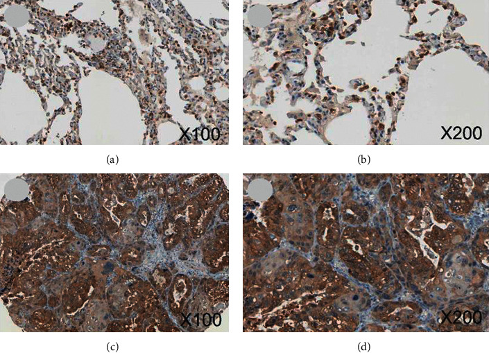
Figure 2.

IHC graphs from HPA. (a, b) ENO1 staining was found in cytoblast of normal alveolar epithelial cells (magnification: 100x and 200x). (c, d) ENO1 was detected in cytoplasmic of LUSC cells, which had stronger staining and stronger intensity (magnification: 100x and 200x). Note: HPA, The Human Protein Atlas; IHC, immunohistochemistry; LUSC, squamous cell carcinoma of lung.
