
Fig. 1 |. High-resolution ELiC structure in a lipid-bound state.
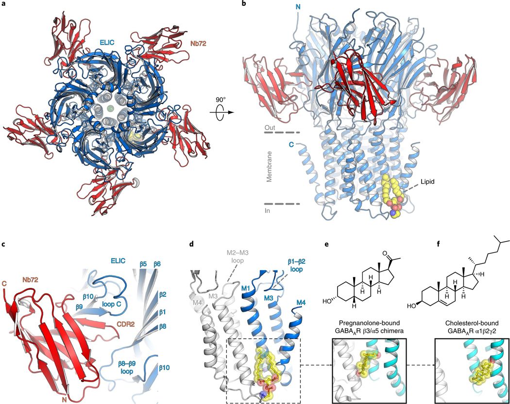
a,b, Top and side views of the crystal structure of ELIC 7′C pore mutant (blue) in complex with nanobody 72 (Nb72, red) in a lipid-bound state. Protein is shown in cartoon representation, lipid in sphere representation. Yellow corresponds to carbon, blue to nitrogen and red to oxygen. c, Detailed view of the interaction between Nb72 and orthosteric binding site in ELIC, including loop C. d, Detailed view of the lipid interaction at a transmembrane site. One subunit is shown in blue and its neighboring subunit in white. PE is shown in transparent sphere and sticks. The lipid site is formed at the M1–M4 interface of one subunit and M3 of the neighboring subunit. This site overlaps with a known neurosteroid binding site in the β3/α5 chimeric GABAA-R29 (e) and a cholesterol-binding site in the α1β2γ2 GABAA-R30 (f).