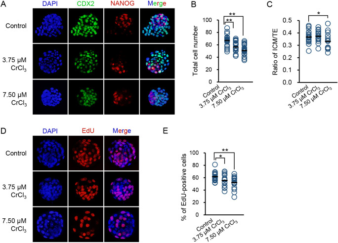
Fig. 3.
Effects of low concentrations of CrCl3 on the quality of blastocysts. (A) Representative images of CDX2, NANOG, and DAPI staining of blastocysts exposed to low concentrations of CrCl3 or no CrCl3. (B and C) Quantification of total cell number (B) and ICM:TE ratio (C) of detected blastocysts in each group. (D) Representative images of EdU staining of blastocysts exposed to low concentrations of CrCl3 or no CrCl3. (E) Quantification of EdU-positive cells of detected blastocysts in each group. * P < 0.05; ** P < 0.01. Three independent experiments were performed.

