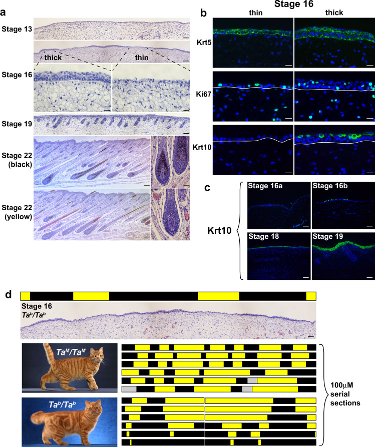
Fig. 1. Patterns of epidermal thickening in fetal cat skin.
a Cat skin histology at different developmental stages (stage 16, bottom panels—high power fields of the thick and thin epidermis; stage 22, right panels—high power fields of black and yellow follicles). b Immunofluorescence for Krt5, Ki67, and Krt10 (green) in stage 16 skin sections (DAPI, blue; white lines mark dermal-epidermal junction). c Krt10 immunofluorescence (green) on cat skin at different developmental stages (DAPI, blue). All micrographs (a) and immunofluorescence (b, c) observations were made independently on three or more embryos with similar results. d Topological maps based on skin histology (thin, yellow; thick, black) from TaM/TaM and Tab/Tab stage 16 embryos mimic pigmentation patterns observed in adult animals (left panels; gray bar—no data). Maps were developed independently on three embryos of each genotype with similar results. Representative images of adult color pattern in TaM/TaM and Tab/Tab animals (from Helmi Flick, with permission) are shown on the left. Scale bars: a stage 13, 50 μM; stage 16 (top), Stage 19, stage 22 (left) 100 μM; stage 16 (bottom), stage 22 (right) 25 μM; b 25 μM; c, d 50 μM.

