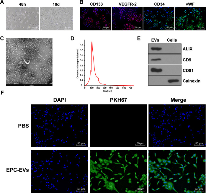
Fig. 1. EPC-EVs could be internalized by endothelial cells.
A The morphology of EPCs was observed under the inverted microscope (bar = 100 μm). B The markers of EPCs were detected using immunofluorescence (CD133 presented red and the nucleus presented blue after Hoechst3342 staining; CD34 presented green and the nucleus presented blue after Hoechst3342 staining; KDR presented red and the nucleus presented blue after Hoechst3342 staining; vWF presented green and the nucleus presented blue after Hoechst3342 staining) (bar = 50 μm). C TEM showed the particle diameter of EPC-EVs (bar = 200 nm). D NTA showed the diameter distribution and concentration of EVs. E The EV marker proteins Alix, CD81, CD9, and endoplasmic reticulum protein calnexin were detected using western blot. F PKH67-labeled EPC-EVs into ECs were observed under the immunofluorescent microscope (PKH67-labeled EPC-EVs presented green and the nucleus presented blue after Hoechst3342 staining) (scale bar = 50 μm). Each experiment was repeated three times independently.

