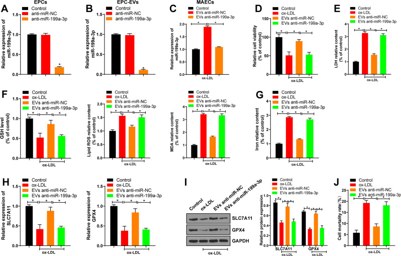
Fig. 3. EPC-EVs inhibited ferroptosis of ECs through miR-199a-3p.
MAECs were isolated from the aorta of C57BL/6J mice and treated with ox-LDL alone or in combination with EVs/EVs-anti miR-199a-3p for 24 h. A The efficiency of lentivirus infection was confirmed using qRT-PCR. B miR-199a-3p expression in EPC-EVs after lentivirus infection was detected using qRT-PCR. C miR-199a-3p expression in MAECs co-cultured with EVs/EVs-anti miR-199a-3p was detected using qRT-PCR. D The viability of MAECs co-cultured with EVs/EVs-anti miR-199a-3p was detected using CCK8 assay. E The cell damage was measured using LDH kit. F GSH consumption, lipid ROS production, and lipid peroxidation were detected using the kits. G The relative content of iron in MAECs was determined using the commercial kit. H, I SLC7A11 and GPX4 mRNA expression and protein level in MAECs were detected. J Cell death rate was detected by trypan blue staining. Each experiment was repeated three times independently. Measurement data are depicted as mean ± SD. The t test was used for the comparisons between two groups. One-way ANOVA was employed for the comparisons among multiple groups, followed by Tukey’s multiple comparisons test. *p < 0.05.

