Fig. 2.
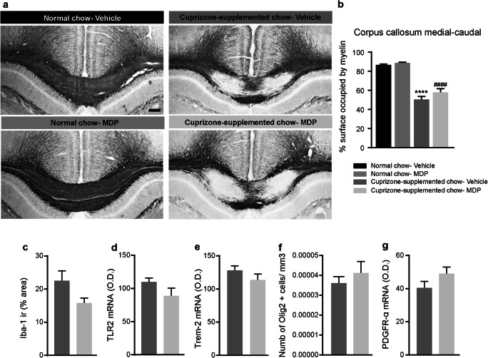
MDP treatment did not significantly modulate remyelination levels, microglia activation, and inflammation in the CNS of cuprizone-fed mice. (a) Representative of Black Gold II staining of the medial-caudal area of the corpus callosum in normal food and cuprizone-supplemented diet. n = 5 mice/group in treatment and control groups in normal food, and n = 10 mice/group in treatment and control groups in cuprizone-supplemented diet. (b) Representative measuring of the medial-caudal area of the corpus callosum occupied by myelin in normal chow (vehicle and MDP) and cuprizone-supplemented chow (vehicle and MDP) groups. Data are expressed as the means ± SEM; ****P < 0.0001 versus normal chow–vehicle, ####P < 0.0001 versus normal chow-MDP, one-way ANOVA with Tukey’s multiple comparisons test. (c) Iba1 was immunostained on the medial-caudal area of the corpus callosum from cuprizone–vehicle and cuprizone–MDP mice. The area covered by Iba1+ staining was measured using a stereological procedure. (d) and (e) In situ hybridization signal of TLR2 and TREM2 mRNA in the medial-caudal area of the corpus callosum from cuprizone–vehicle and cuprizone–MDP mice. (f) Representative number of Olig2-immunoreactive staining (olig2+ cell/μm3) in medial–caudal area of the corpus callosum from cuprizone–vehicle and cuprizone–MDP mice. (g) PDGFRα mRNA hybridization signal in the medial-caudal area of the corpus callosum of cuprizone–vehicle and cuprizone–MDP mice

