Fig. 4.
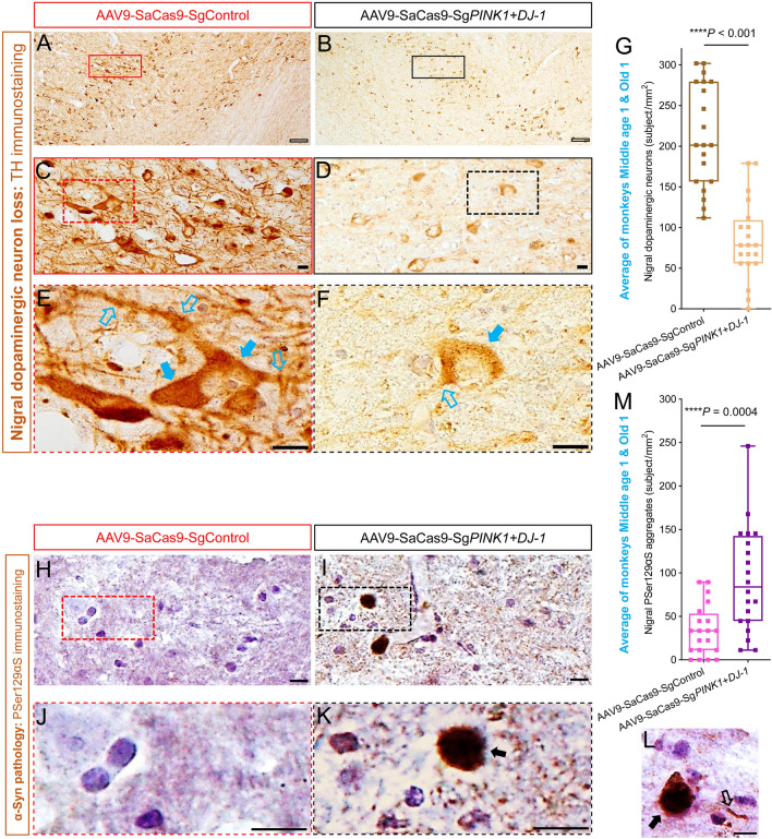
Pathological hallmarks of PD in the SN region of PINK1 and DJ-1 gene-edited monkey brain. A, B TH immunostaining images of the AAV9-SaCas9-SgControl SN (A) and the AAV9-SaCas9-SgPINK1+DJ-1 SN (B) of monkey Old 1 at low resolution (4×). The image in B appears pale and weak with fewer surviving neurons compared with A. C, D Moderate resolution (20×) images of boxed areas in A and B. Typical dopaminergic neurons are evident in C, and many deformed dopaminergic neurons, with weakly-stained somata and almost completely lost neurites are shown in D. E, F High resolution (40×) images of the boxed areas in C and D. A typical dopaminergic neuron with intact soma (solid blue arrows) and branched processes (hollow blue arrows) can be seen in E, and a very weakly-stained dopaminergic neuron with incomplete soma boundary (solid blue arrow) and a residual process (hollow blue arrow) is shown in F. G Over 61% of nigral dopaminergic neurons are lost in AAV9-SaCas9-SgPINK1+DJ-1 on the edited side compared with the AAV9-SaCas9-SgControl side (an average of monkeys Old 1 and Middle age 1). H, I PSer129αS immunostaining images under high resolution (40×) of the AAV9-SaCas9-SgControl SN (H) and the AAV9-SaCas9-SgPINK1+DJ-1 SN (I) from monkey Old 1. Clear PSer129αS aggregates can be seen in I. J, K Enlarged images of the defined areas in H and I showing stained nuclei in H and a typical darkly-stained PSer129αS aggregate in I (arrow). L Typical example of a PSer129αS aggregate (solid arrow) and a PSer129αS deposit in a neuronal process (hollow arrow) in the AAV9-SaCas9-SgPINK1+DJ-1 SN of monkey Old 1. M Numbers of PSer129αS aggregates in the AAV9-SaCas9-SgPINK1+DJ-1 and the AAV9-SaCas9-SgControl SNs (average of monkeys Old 1 and Middle age 1). Scale bars: hollow black, 100 μm; solid black, 10 μm. Data in G and M are the median with minimum and maximum (n = 20 sections).

