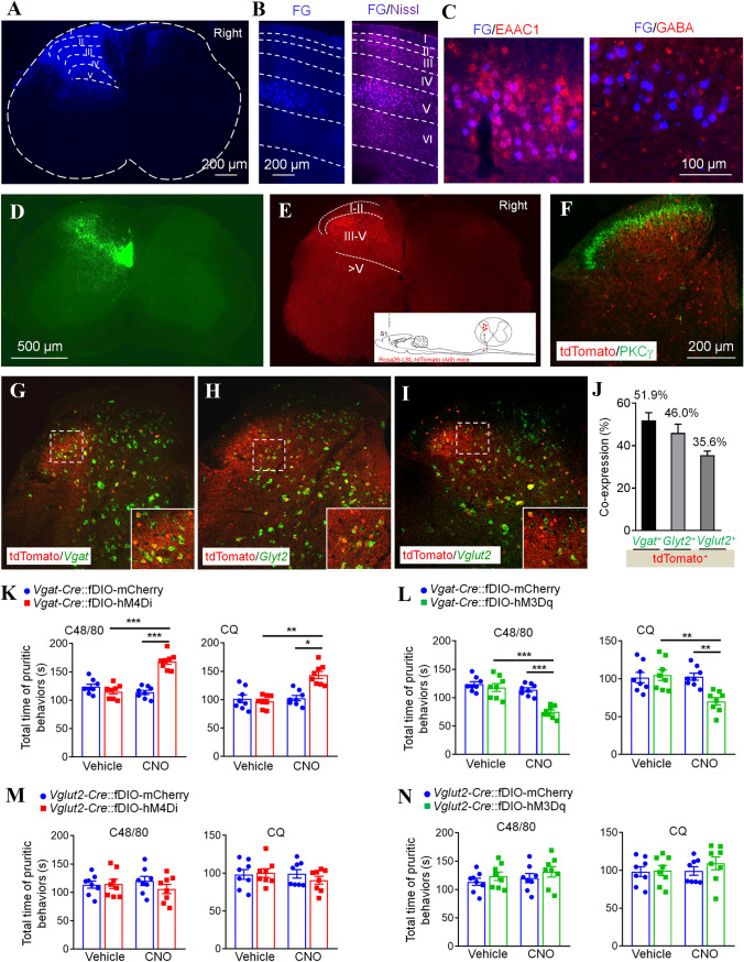
Fig. 2.
Inhibitory interneurons of the spinal dorsal horn mediate the inhibitory effect of S1-SDH projection on itch transmission. A Fluorescence image showing a site of FG injection in the spinal dorsal horn. B Coronal brain section showing the location of FG+ neurons in the S1. C Immunostaining against EAAC1 or GABA with FG in the S1. D Image showing the S1-SDH tract terminates in the deep laminae of the spinal dorsal horn. E Representative image showing tdTomato+ post-synaptic neurons of the S1-SDH projection in the spinal dorsal horn. The insert shows the strategy for identifying spinal post-synaptic neurons of the S1-SDH projection. F Representative image showing the staining of PKCγ with tdTomato in the spinal dorsal horn. G–I In situ hybridization showing Vgat (G), Gylt2 (H), and Vglut2 (I) with tdTomato in spinal sections, and statistical data showing the percentages of tdTomato+ neuronal co-expression with Vgat, Glyt2, and Vglut2 (J). K Pharmacogenetic inhibition of spinal post-synaptic Vgat+ neurons enhances itch behaviors in response to C48/80 or CQ (n = 8). L Pharmacogenetic activation of spinal post-synaptic Vgat+ neurons alleviates itch behaviors in response to C48/80 or CQ (n = 8). M Pharmacogenetic inhibition of spinal post-synaptic Vglut2+ neurons has no effect on itch behaviors in response to C48/80 or CQ (n = 8). N Pharmacogenetic activation of spinal post-synaptic Vglut2+ neurons has no effect on itch behaviors in response to C48/80 or CQ (n = 8).

