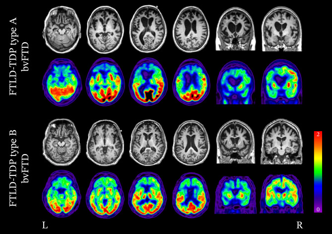
Fig. 2.
Neuroimaging of bvFTD associated with FTLD-TDP type A and B (top row: structural MRI; bottom row: FDG-PET). The variability in patterns of degeneration attributed to FTLD-TDP neuropathology is readily seen in these two cases of bvFTD. In the case of bvFTD associated with FTLD-TDP type A, degeneration and hypometabolism of the bilateral frontal lobes are present; however, the left is significantly more impacted than the right and extends to the left parietal lobe. In bvFTD associated with FTLD-TDP type B, significant degeneration of the bilateral frontal lobes is seen; however, in contrast to type A, the parietal lobes are less affected and atrophy is relatively symmetrical

