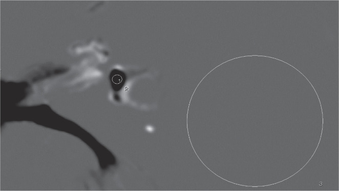
Fig. 1.
An example of ROI placement. Circular ROI #1 is drawn in the vestibular endolymph as large as possible on the iHYDROPS-Mi2 image. Circular ROI #2 is drawn in the perilymph as large as possible. Circular ROI #3 is drawn as a circle with the diameter of 20 mm in the area of uniform signal intensity that is lateral to the labyrinth for the noise level estimation. The contrast-to-noise ratio between the perilymph and the endolymph is defined as a subtraction of the signal intensity of ROI #2 minus ROI #1 divided by the standard deviation of ROI #3. iHYDROPS-Mi2, improved hybrid of reversed image of the positive endolymph signal and the native image of the perilymph signal multiplied with the heavily T2-weighted MR cisternography; ROI, region of interest.

