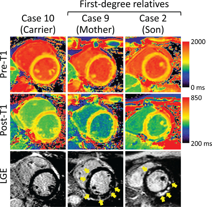
Fig. 1.
Representative images in a putative carrier of Becker muscular dystrophy (Case 10), a patient with Duchenne muscular dystrophy (Case 2), and the mother of Case 2 (Case 9; a putative carrier of Duchenne muscular dystrophy). Case 2 and 9 show nonischemic patchy hyperenhancement (yellow arrows) on the LGE image and abnormal pre- and post-contrast T1 values located in the same areas. Both putative carriers had an elevated extracellular volume fraction (37.4% and 36.1%, respectively). LGE, late gadolinium enhancement.

