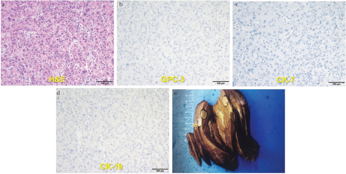
Fig. 3.
Histopathologic and immunohistochemical photomicrographs (×200) of one patient whose MR images are displayed in Fig. 1. (a) H&E stained section (low grade, without MVI), (b) GPC-3 stained section (GPC-3 expression: negative), (c) CK-7 stained section (CK-7 expression: negative), (d) CK-19 stained section (CK-19 expression: negative) and (e) surgical specimen (Note: There was no micro-hemorrhage according to the specimen.). CK-7, cytokeratin 7; CK-19, cytokeratin 19; GPC-3, Glypican-3; MVI, microvascular invasion.

