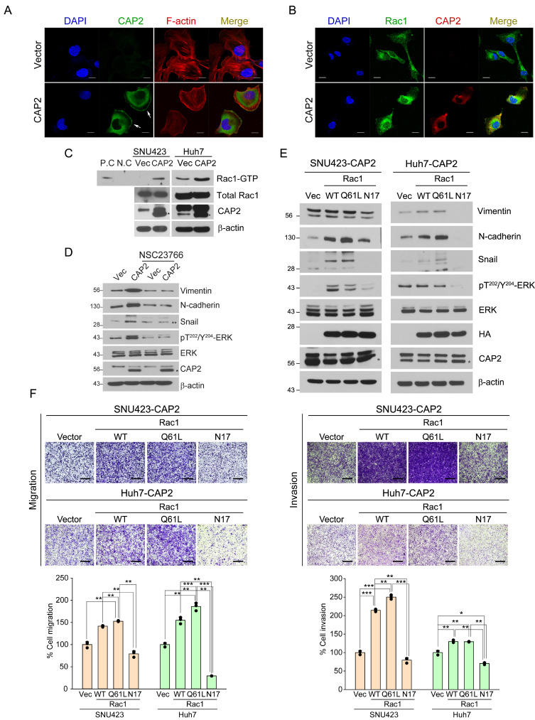
Fig. 5. Rac1 is involved in CAP2-mediated ERK activation and EMT.
(A and B) SNU423-Vector and SNU423-CAP2 cells are stained with the indicated fluorescent antibodies. Nuclei are counter-stained with DAPI. Images are captured under a laser scanning confocal microscope (×40). White arrows indicate membrane ruffles. Scale bars = 10 μm. (C) Total cell lysates prepared from the indicated cells are incubated with agarose beads coupled to PAK PBD. Bound Rac1 is detected with Western blotting using a Rac1 Ab (upper panel). The amount of total Rac1 is also measured (lower panel). P.C, positive control; N.C, negative control. Asterisks (*) indicate CAP2. (D) SNU423-Vector and SNU423-CAP2 cells were treated for 24 h with NSC23766 (50 μM) and subjected to western blotting with the indicated antibodies. Asterisk (*) indicates CAP2, double asterisk (**) indicates Snail. (E) SNU423-CAP2 and Huh7-CAP2 cells stably expressing Vector (Vec), Rac1-WT, Rac1-Q61L, or Rac1-N17 are subjected to Western blotting with the indicated antibodies. Asterisks (*) indicate CAP2. (F) Migration and invasion of the indicated cells were measured in Transwell assays. Migrated/invaded cells are fixed with 10% formaldehyde and stained with 1% crystal violet. WT, wild type. Images are captured under a light microscope (magnification, ×5). Scale bars = 500 μm. Data are presented as mean ± SEM for triplicate experiments. *P < 0.05, **P < 0.01, and ***P < 0.001.

