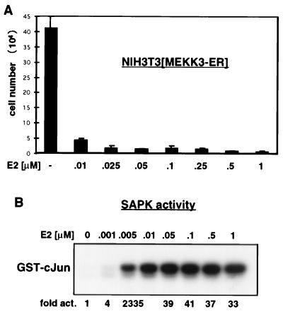
FIG. 2.
Activation of MEKK3 inhibits proliferation. (A) MEKK3-ER prevents cell division. NIH 3T3 [MEKK3-ER] cells were seeded in triplicate at 2 × 10⁴ per well of a six-well plate in complete medium in the absence or presence of increasing concentrations of E2, as indicated. After 4 days, the attached cells were harvested by trypsinization, pooled with the detached cells, and counted in the presence of trypan blue to distinguish between live and dead cells. The percentage of dead cells was marginal, even with the highest concentration of E2. The total number of cells counted is indicated on the y axis (±mean standard deviation). As discussed in Results, no significant cell death could be measured. (B) Activation of SAPK by MEKK3-ER in the presence of increasing concentrations of E2. NIH 3T3 [MEKK3-ER] cells were transiently transfected with HA-tagged SAPK, serum starved overnight, and stimulated for 15 min with the indicated concentrations of E2. The activity of SAPK was measured in an immunocomplex kinase assay with GST–c-Jun as the substrate. After SDS-PAGE, phosphorylated GST–c-Jun was quantified with a Phosphorimager; data are expressed as fold activation relative to the signal obtained in the absence of E2. Western blot analysis confirmed equal expression of HA-SAPK in the different extracts (data not shown). Activation of SAPK and growth inhibition appear to occur at roughly the same low doses of E2.

