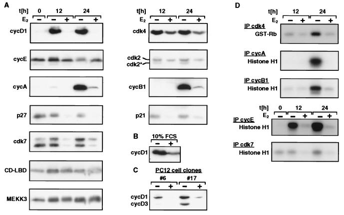
FIG. 4.
Activation of MEKK3 inhibits various molecular parameters of cell cycle progression, as determined by analysis of inhibition of cyclin expression and CAK activities. (A and D) NIH 3T3 [MEKK3-ER] cells were kept without serum for 4 days in the presence or absence of E2 and then stimulated with 10% FCS for 12 or 24 h in the presence or absence of E2, as indicated. For the Western blot shown in panel B, cells were continuously kept in medium containing 10% FCS in the presence or absence of E2. cycD1, cyclin D1. (C) Two different clones of PC12 cells stably expressing MEKK3-ER (#6 and #17) were starved in the absence of FCS for 1 day with or without E2 and then released into medium containing 10% FCS for 2 days (with or without E2), as indicated. (A to C) Detergent lysates (100 μg) were electrophoretically separated on denaturing polyacrylamide gels and immunoblotted with antibodies against the proteins indicated on the left. CD-LBD designates the MEKK3-ER fusion protein, and cdk2* designates the T160 phosphorylated form of cdk2. (D) An aliquot of 100 μg (for cdk4 kinase assay) or 50 μg (all others) of extract was immunoprecipitated (IP) with the indicated antibodies and then assayed for associated kinase activity in an immunocomplex kinase assay with histone H1 or GST-pRb as the substrate.

