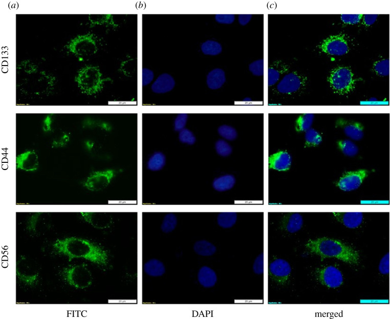
Figure 3.
Immuno fluorescent lung CSC characterization. Fluorescent antigenic detection of the surface markers CD133, CD44 and CD56. (a) Immunofluorescent staining of the isolated side population of lung CSCs using FITC. (b) Cells counterstained with DAPI indicated by blue FL seen in the nuclei. (c) Superimposed fluorescent images of the labelled cells.

