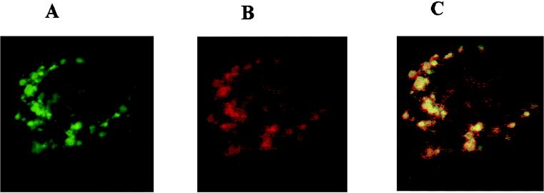
FIG. 3.
Colocalization within the mitochondria of GFP autofluorescence and rhodamine-labeled anti-GFP antibody. Panels show immunofluorescence of Cos-1 cells transfected with plasmid pLIGIII-GFP-2. (A) GFP fluorescence; (B) cells incubated with anti-GFP monoclonal antibody and then stained with rhodamine labeled anti-mouse immunoglobulin G antibody; (C) merged images of GFP and rhodamine fluorescence from panels A and B.

