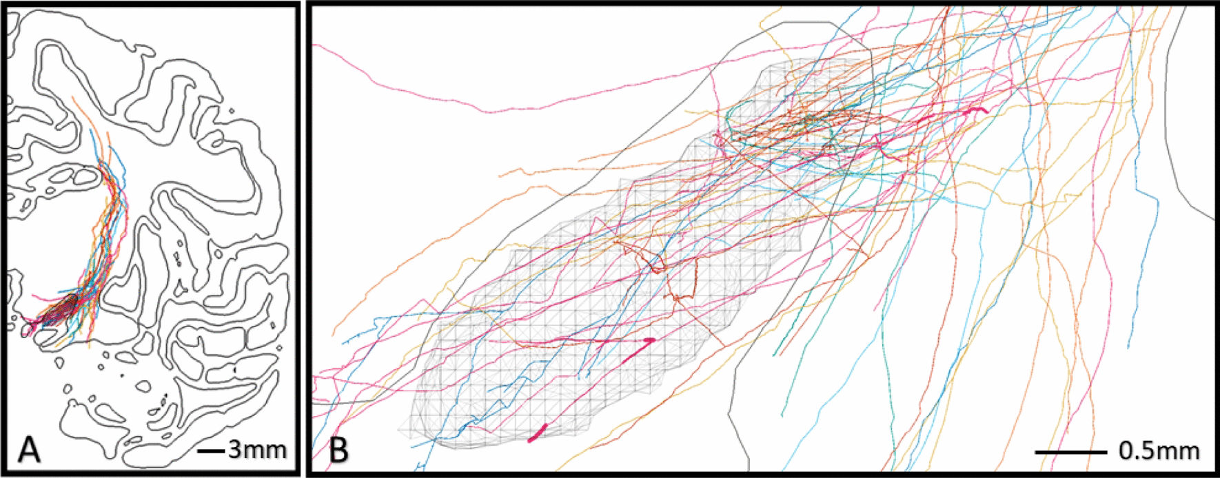
Figure 4.

Explicit motor HDP reconstructions in the STN. Visualization of 24 HDP axons registered with the 3D cynomolgus brain atlas. A) Cut-plane view of the left hemisphere showing organization of axons within the internal capsule and collateralizations which principally target the STN (hatched volume). B) Topography of collaterals within the subthalamic region shows a focus on the dorsal STN, but axon collaterals also innervate the medial and ventral regions of the STN.
