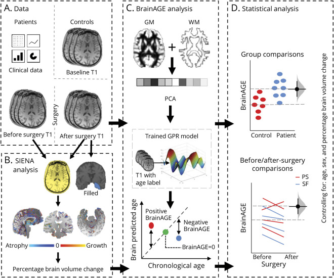
Figure 1. Graphic Description of the Analysis Pipeline.
(A) In addition to clinical patient data, 3D T1-weighted MRI scans were acquired for 48 patients with mesial temporal lobe epilepsy before and after temporal lobe surgery and for 37 controls. (C) Brain age was estimated for each scan using a trained gaussian processes regression (GPR) model following tissue segmentation, vectorization, and principal components analysis (PCA)–based dimension reduction. Brain age gap estimation (BrainAGE) was computed as brain-predicted age minus actual age. BrainAGE comparisons were made between patients and control groups before and after epilepsy surgery, controlling for percentage brain volume change (SIENA; B), age, and sex (D). GM = gray matter; WM = white matter.

