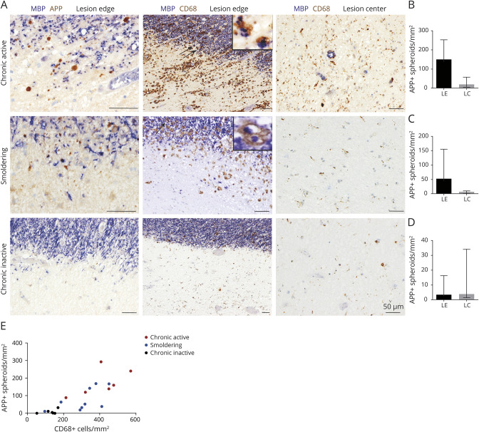
Figure 3. Acute Axonal Damage in Chronic Active, Smoldering, and Chronic Inactive Lesions.
(A) Double immunohistochemistry for myelin basic protein (MBP) (blue) with human β-amyloid precursor protein (APP) (brown) or CD68 (clone KiM1P) at the lesion edge and center of chronic active, smoldering, and chronic inactive lesions. Insets highlight CD68+ phagocytes containing MBP+ particles. (B–D) Quantification of APP+ spheroids at the lesion edge (LE) and center (LC) of chronic active lesions (n = 6; Mann-Whitney U test, p = 0.004), smoldering lesions (n = 9; Mann-Whitney U test, p = 0.0002), and chronic inactive lesions (n = 6; Mann-Whitney U test, p = 0.46). (E) Correlation of the density of APP + spheroids with the density of CD68+ macrophages/activated microglia at the lesion border of chronic active/smoldering and chronic inactive lesions (n = 21; r = 0.8648; p < 0.0001; nonparametric Spearman correlation). CD68 = CD68-macrophage/activated microglia antibody (clone KiM1P).

