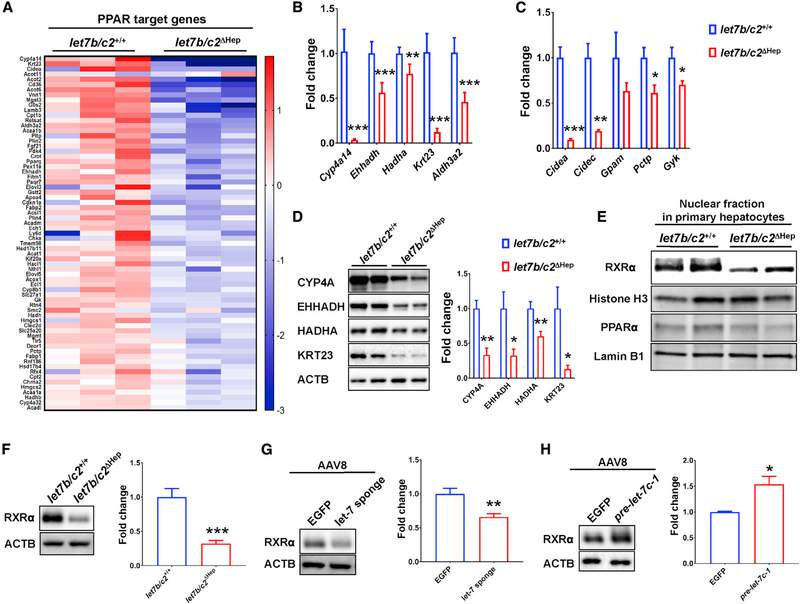
Figure 3. PPARα target gene expressions were repressed by RXRα protein reduction in let7b/c2ΔHep and let-7 sponge AAV-transduced mice.
(A) Heatmap of PPARα target genes identified by differential gene expression analysis of RNA-seq data from let7b/c2+/+ and let7b/c2ΔHep livers after HFD feeding.
(B and C) mRNA analysis by qRT-PCR of PPARα target genes involved in fatty acid oxidation and cell proliferation (B) and lipid accumulation and glucose metabolism (C) in HFD-fed let7b/c2+/+ and let7b/c2ΔHep livers.
(D) Western blot analysis for PPARα target genes in HFD-fed let7b/c2+/+ and let7b/c2ΔHep liver lysates.
(E) Western blot analysis of PPARα and RXRα protein expression in nuclear fractions isolated from let7b/c2+/+ and let7b/c2ΔHep hepatocytes.
(F–H) Western blot analysis of RXRα and the densiometric quantification in whole-liver lysates from let7b/c2ΔHep- mice (F), let-7 sponge expressing AAVinflected mice (G), and pre-let-7c-1--AAVinfected mice (H).
Data are presented as mean ± SEM (n = 4–5 mice per group; *p < 0.05, **p < 0.01, ***p < 0.001

