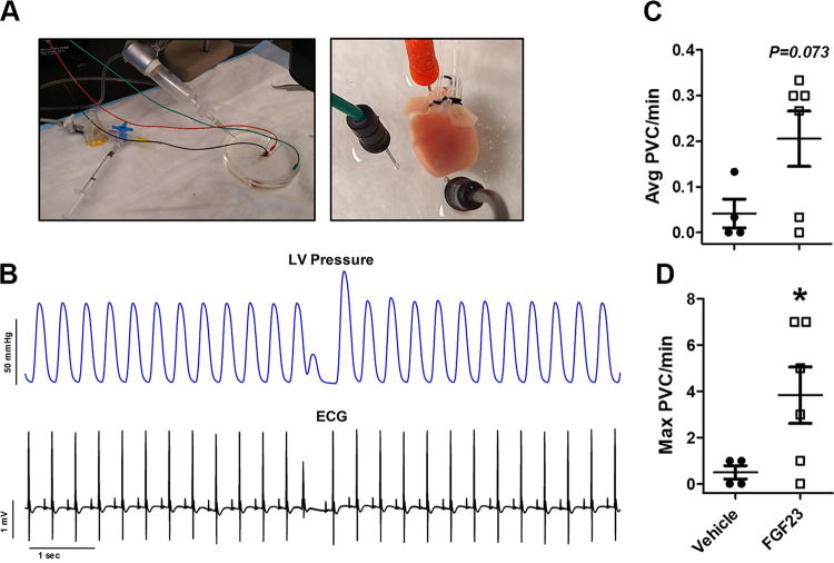
Figure 2.
A: images of the ex vivo electrocardiogram (ECG) and left ventricular (LV) pressure setup. Electrodes were placed at the base and apex of the heart (along with a ground wire) in the dish to measure ECG during Langendorff perfusion while a balloon cannula was inserted into the LV via the left atria to measure pressure changes. B: ventricular dysrhythmias after fibroblast growth factor 23 (FGF23) (9 ng/mL) perfusion were confirmed via ex vivo ECG synchronized with intraventricular pressure via the LV balloon catheter. Average premature ventricular contractions (PVCs) per min (C) and maximal number of PVCs per minute by ECG after 30 min of perfusion with vehicle (n = 4) or FGF23 (n = 6) (D). *P < 0.05 compared with vehicle.

