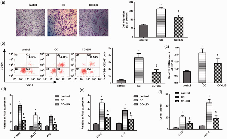
Figure 2.
The ability of HCC cells to induce macrophage recruitment and M2 polarization was abrogated by ligustilide. (a) HCC cells under ligustilide exposure, or not, were co-cultured with PMA-treated THP-1 macrophages using the Transwell co-culture system. Macrophage migration was then analyzed. Macrophages without any treatment were defined as a control group. Scale bars, 100 µM. (b) Flow cytometry was used to detect the percentage of CD14+CD206+ cells in macrophages. (c and d) The mRNA levels of M2 macrophage phenotype markers were determined by qRT-PCR. (e and f) The transcript (e) and release (f) of M2 macrophage marker IL-10 and TGF-β were further explored. *P < 0.05 vs. control group. §P < 0.05 vs. CC-treated group. (A color version of this figure is available in the online journal.)

