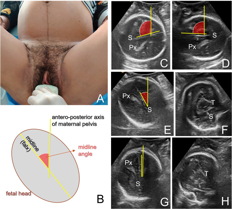
Figure 3.
Ultrasound determination of the occiput position in the second stage of labour. (A) Example of the probe orientation in the transperineal infrapubic transverse plane. (B) Schematisation of the structures visualised and the measurement of the midline angle between the midline (falx cerebri) and the anteroposterior axis of the maternal pelvis. (C–H) Presentation of an example with midline angle measurement and evolution during anterior rotation of a transverse occiput. Occiput position is identified based on the visualisation of the cerebral midline (interhemispheric septum, (S) and choroid plexus (Px) direction (divergent posteriorly), or thalami aspect (triangular, with the base anteriorly). Midline angle gradually decreases during the anterior occiput rotation from the transverse position (C, D), as it reaches right anterior (E, F) and anterior (infrapubic) (G, H) positions.

