FIG. 3.
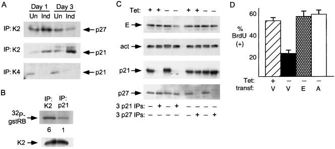
Inhibition of cdk2 by p21 in cells with p16 induction. (A) Increased association of p21 with cdk2 and decreased association with cdk4 following p16 induction. Extracts were prepared from cells cultured with or without p16 induction for 1 and 3 days and were normalized for protein content. cdk2 (K2) or cdk4 (K4) complexes were immunoprecipitated and probed for coprecipitated p21 or p27 by immunoblotting. Un, uninduced; Ind, induced. (B) Inhibition of cdk2 kinase activity in p21-bound complexes. Extracts were prepared from cells with p16 induction for 3 days, and p21 and cdk2 were immunoprecipitated. The immunoprecipitates were normalized for cdk2 protein level and assayed for cdk2 activity with gstRB as a substrate. Quantitation of the relative band intensity is provided below each lane. (C) Immunodepletion of cyclin E bound to p21 following 3 days of p16 induction. Extracts were prepared from cells with (+) or without (−) TET for 3 days and subjected to three successive immunoprecipitations with antibodies directed against p21 or p27. Cyclin E, actin (act; a negative control), p21, and p27 levels in the starting extracts and in the supernatant following the third IP were assessed by immunoblotting. Clearing of a major fraction of cyclin E from the supernatant occurred only following p21 IP, and only in extracts prepared from cells with p16 induction in this and two other experiments (data not shown). Parallel IPs with a negative-control antiserum (anti-hemagglutinin [12CA5]) had no effect on the level of any of these antigens (data not shown). (D) Overexpression of cyclin E or A can prevent the arrest mediated by p16. OSp16.1 cells were cotransfected with a CMV vector expressing β-gal together with an empty expression vector (V) or one expressing cyclin E (E) or cyclin A (A). A portion of the vector-transfected (transf.) cells was replated in the presence of TET as a positive control for BrdU incorporation. p16 was induced in the remaining cells by replating in the absence of TET for 24 h. The cells were then incubated with BrdU for 8 h, fixed, and stained for β-gal and BrdU. One hundred β-gal-positive cells per condition were scored by an observer blinded to the treatment groups, and the results confirmed by a second, similarly blinded observer. The mean percentage of BrdU-positive cells from three experiments is depicted. Error bars indicate standard deviations.

