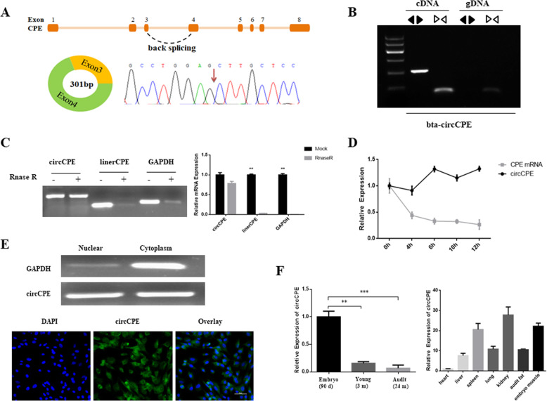
Fig. 1.
Characterization of bovine CircCPE. (A) Schematic showing the circularization of CPE exon 3 and exon 4 forming circCPE. The back- splicing junction of circCPE was confirmed by Sanger sequencing. (B) The existence of circCPE was verified with PCR and agarose gel electrophoresis assay using divergent primers and convergent primers. (C) Resistance to RNase R of circCPE was tested using agarose gel electrophoresis and qRT-PCR assay (D) qRT-PCR for the abundance of circCPE and CPE mRNA in myoblasts treated with Actinomycin D at the indicated time points, and the results indicated that circCPE was more stable than linear CPE. (E) Cellular localization analysis and the RNA-FISH assay showed that circCPE is localized both in the cytoplasm and nuclear. GAPDH is positive control for cytoplasmic fraction. Blue indicates nuclei stained with DAPI; green indicates the RNA probe that recognizes circCPE. (F) Expression profile of circCPE in muscle tissues from fetal to adult cattle and in different tissues of fetal cattle. Data are presented as means ± SEM. P < 0.05, P < 0.01

